Myeloma Novel Drug Targets and agents
Poster Session 2
P-187: Therapeutically Targeting 19S Proteasome-associated Subunit PSMD1/Rpn2 in Multiple Myeloma
Thursday, September 28, 2023
12:30 PM - 1:30 PM EEST


Ting Du, PhD (she/her/hers)
Instructor
Dana-Farber Cancer Institute
BOSTON, Massachusetts, United States
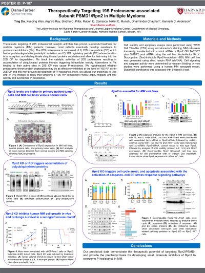
Introduction: Therapeutic targeting of 20S proteasomal catalytic activities has proven successful treatment for multiple myeloma (MM) patients; however, most patients eventually develop resistance to proteasome inhibitors (PIs). The 26S proteosome is composed of 1) 20S core particle (CP) which harbor protein-degradative proteolytic activities, and 2) 19S regulatory particle (RP) whose function is to recognize, and deubiquitinate ubiquitinated proteins substrates and allow for their entry into the 20S CP for degradation. PIs block the catalytic activities of 20S proteasome resulting in accumulation of ubiquitinated proteins thereby triggering intracellular toxicity. Aberrations in PIs binding to their active sites in 20S CP may cause PI-resistance. We hypothesized whether proteasome-driven protein degradation may be as effectively inhibited at the level of 19S RP as for 20S CP and this may prevent development of PI-resistance. Here, we utilized our preclinical in vitro and in vivo models to show that targeting a 19S RP component PSMD1/Rpn2 triggers anti-MM activity and overcomes PI-resistance.
Methods: Cell viability was determined by CellTiter-Glo Luminescent assays (Promega Corporation). Apoptosis was measured using Annexin/PI staining (Biolegend). MM cells were transiently transfected with Rpn2 siRNA using the Nucleofector Kit V. Dox-inducible Rpn2-KD MM cell line was generated using shRNA. In vivo studies were performed using a human MM xenograft model. Statistical significance was assessed with Student’s t test.
Results: 1) Immunoblot analysis showed higher Rpn2 expression in MM vs normal peripheral blood mononuclear cells. 2) Immunohistochemistry studies showed higher Rpn2 expression in BM biopsies from MM patients than from healthy individuals. 3) DepMap analysis showed that Rpn2 was essential for MM cell lines. 4) Knocking down (KD) Rpn2 using transiently transfected siRNA decreased the viability of various MM cell lines, including those that are PI-resistant (ANBL6.BR) or carry p53 alterations (JJN3, ARP1) (p < 0.01). 5) Transfection with Rpn2-WT rescued cells from the growth-inhibitory activity of Rpn2-siRNA. 6) Dox-inducible CRISPR/Cas9 stable Rpn2-KD and -KO MM cells reduced cell growth. 7) Mechanistically, Rpn2 blockade both inhibited proteasome-mediated protein degradation, evident by increased ubiquitinated proteins levels, and triggered cell death/apoptosis associated with the activation of caspases, and endoplasmic reticulum stress response signaling. 8) Proteomic analysis showed decreased cell-cycle- and DNA replication-related pathway proteins in Rpn2 KD vs Rpn2 -WT cells. Finally, 9) Using Dox-inducible Rpn2-KD AMO1 MM cells in a xenograft mouse model, we found that Rpn2 depletion reduced tumor growth and prolonged mouse survival (p < 0.005).
Conclusions: Our preclinical data demonstrate the therapeutic potential of targeting Rpn2 and provide the preclinical basis for developing Rpn2 inhibitors to overcome PI resistance in MM.
