Imaging and Management of Myeloma Bone Disease
Poster Session 1
P-063: Comparison of whole-body imaging in multiple myeloma with respect to lytic involvement and presence of extramedullary disease
Wednesday, September 27, 2023
1:30 PM - 2:30 PM EEST


Jiri Minarik, MD, PhD (he/him/his)
physician
University Hospital Olomouc
Olomouc, Olomoucky kraj, Czech Republic
Introduction: The presence of osteolytic involvement in multiple myeloma (MM) is one of the halmarks of the disease. In approximately 5-20% of patients, prognostically negative paramedullary and/or extramedullary disease (EM) is detectable, based on the imaging technique used.
Conventional X-ray has only limited sensitivity in the detection of lytic involvement, especially in the spine, and a very low sensitivity for EM. Therefore, more sophisticated whole-body imaging techniques are recommended, such as magnetic resonance imaging (MRI), low dose computed tomography (LD-CT) or positron emission tomography with computed tomography (PET/CT). Each of them has slightly different outcomes and limitations.
Our aim was to compare the sensitivity of selected whole-body imaging techniques in the detection of osteolytic involvement and EM.
Methods: We performed a large retrospective assessment of 411 MRI, 304 PET/CT, 269 Technetium-sestaMIBI and 151 LD-CT findings in 270 patients with MM, smoldering myeloma (SMM) and monoclonal gammopathy of undetermined significance (MGUS). We assessed the presence and number of lytic lesions by each technique and similarly the presence and number of EM. In 80 patients we had concurrent data from PET/CT, MRI and MIBI at the same time, therefore we compared these patients and sensitivity of the three techniques separately.
We used statistical software IBM SPSS Statistics, version 23. For the comparison of the techniques, we used Chi-quadrat test, McNemara test, Friedman test and Wilcoxon pair test with Bonferroni correction, the level of statistical significance was p ˂ 0,05.
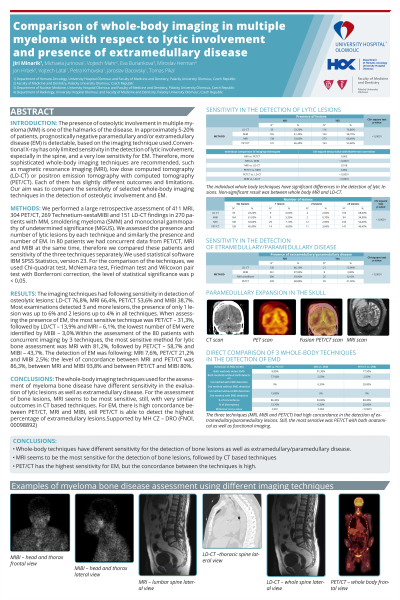
Results: The imaging techniques had following sensitivity in detection of osteolytic lesions: LD-CT 76,8%, MRI 66,4%, PET/CT 53,6% and MIBI 38,7%. Most examinations detected 3 and more lesions, the presence of only 1 lesion was up to 6% and 2 lesions up to 4% in all techniques. When assessing the presence of EM, the most sensitive technique was PET/CT – 31,3%, followed by LD/CT – 13,9% and MRI – 6,1%, the lowest number of EM were identified by MIBI – 3,0%.
Within the assessment of the 80 patients with concurrent imaging by 3 techniques, the most sensitive method for lytic bone assessment was MRI with 81,2%, followed by PET/CT – 58,7% and MIBI – 43,7%. The detection of EM was following: MRI 7,6%, PET/CT 21,2% and MIBI 2,5%; the level of concordance between MRI and PET/CT was 86,3%, between MRI and MIBI 93,8% and between PET/CT and MIBI 80%.
Conclusions: The whole-body imaging techniques used for the assessment of myeloma bone disease have different sensitivity in the evaluation of lytic lesions as well as extramedullary disease. For the assessment of bone lesions, MRI seems to be most sensitive, still, with very similar outcomes in CT based techniques. For EM, there is high concordance between PET/CT, MRI and MIBI, still PET/CT is able to detect the highest percentage of extramedullary lesions.
Supported by MH CZ – DRO (FNOl, 00098892)
