Imaging and Management of Myeloma Bone Disease
Poster Session 1
P-059: Integrative radiomic and immunological analysis for risk classification and outcome prediction in monoclonal gammopathies
Wednesday, September 27, 2023
1:30 PM - 2:30 PM EEST

.jpg)
Emilia Gigliotta, MD (she/her/hers)
physician
Policlinico Universitario "P. Giaccone", Palermo, Italy
Palermo, Sicilia, Italy
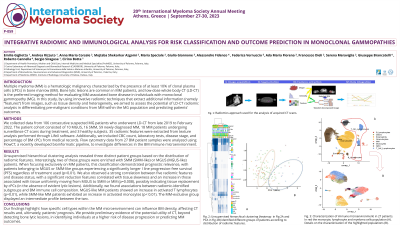
Introduction: Multiple myeloma (MM) is a hematologic malignancy characterized by the presence of at least 10% of clonal plasma cells (cPCs) in bone marrow (BM). Bone lytic lesions are common in MM patients, and low-dose whole body CT (LD-CT) is the preferred imaging method for evaluating MM-associated bone disease in individuals with monoclonal gammopathy (MG). In this study, by using innovative radiomic techniques that extract additional information (namely “features”) from images, such as tissue density and heterogeneity, we aimed to assess the potential of LD-CT radiomic analysis in differentiating pre-malignant conditions from MM within the MG population and predicting patients' outcomes.
Methods: We collected data from 106 consecutive suspected MG patients who underwent LD-CT from late 2019 to February 2022. The patient cohort consisted of 10 MGUS, 16 SMM, 59 newly diagnosed MM, 18 MM patients undergoing surveillance CT scans during treatment, and 3 healthy subjects. 35 radiomic features were extracted from texture analysis performed through LifeX software. Additionally, we included CBC count, laboratory tests, disease stage, and percentage of BM cPCs from medical records. Flow cytometry data from 27 BM patient samples were analyzed using FlowCT, a recently developed bioinformatic pipeline, to investigate differences in the BM immune microenvironment.
Results: Unsupervised hierarchical clustering analysis revealed three distinct patient groups based on the distribution of radiomic features. Interestingly, two of these groups were enriched with SMM (SMM-like) or MGUS (MGUS-like) patients. When focusing exclusively on MM patients, this classification demonstrated prognostic relevance, with patients belonging to MGUS or SMM-like groups experiencing a significantly longer I line progression-free survival (PFS) regardless of treatment used (p=0.01). We also observed a strong correlation between five radiomic features and disease status, with a significant reduction features correlated with tissue skewness and an increase in those associated with tissue uniformity moving from MGUS to SMM or MM (p=0.008), possibly indicating tissue replacement by cPCs (in the absence of evident lytic lesions). Additionally, we found associations between radiomic-identified subgroups and BM immune cell composition. MGUS-like MM patients showed an increase in activated T lymphocytes (p=0.01), while SMM-like MM patients exhibited an increase in activated monocytes (p < 0.01). The MM-exclusive group displayed an intermediate profile between the two.
Conclusions: Our findings highlight how specific cell types within the MM microenvironment can influence BM density, affecting CT results and, ultimately, patients' prognosis. We provide preliminary evidence of the potential utility of CT, beyond detecting bone lytic lesions, in identifying individuals at a higher risk of disease progression or predicting MM outcomes.
